Maud /
DataCoverageInAxialDiffractionExperiments
< Using pole figures to represent directions in space | Radial Diffraction | Data Coverage In Radial Diffraction Experiments >
If you perform an axial diffraction experiment, your incoming x-ray beam is parallel to the compression direction. In the figure below, we show the location of the data measured at eta=0 and eta=90 on the pole figure of the compression direction. Eta is the azimuth angle on the image plate. I personaly call it delta in my publications, but MAUD calls it eta...
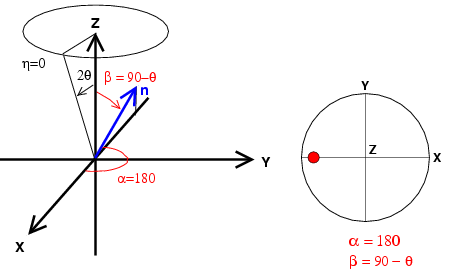 | Figure 1: diffraction in axial geometry with the azimuthal angle on the image plate equals to 0. n is the normal to the diffraction plane and we report the orientation of n on the pole figure. |
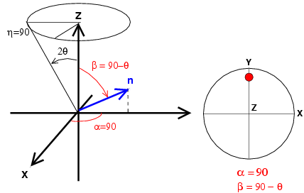 | Figure 2: diffraction in axial geometry with the azimuthal angle on the image plate equals to 90. n is the normal to the diffraction plane and we report the orientation of n on the pole figure. |
Overall, on a pole figure of the compression direction, one diffraction ring will provide you with the following coverage
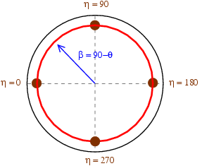 | Figure 2: coverage for diffraction experiments in the axial geometry. Diffractions corresponding to azimuth angles 0, 90, 180, and 360 degrees on the image plate are located in brown on the figure. |